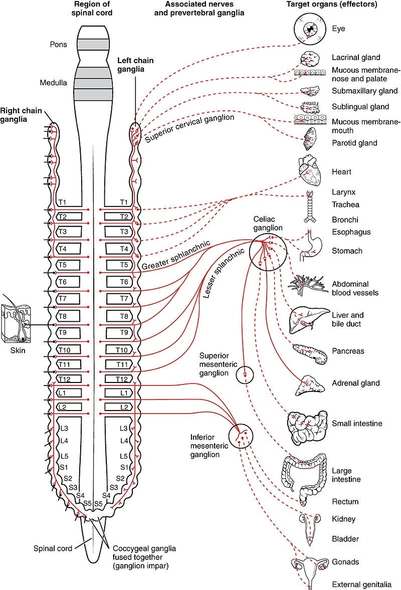
The sympathetic division is involved in the fight, flight, fright, and sex response.
Cell bodies of the sympathetic preganglionic neurons are located in the lateral horn of the spinal cord gray matter between T1-L2.
Their axons traverse the ventral horn to exit in ventral roots where they form synapses onto many postganglionic neurons, thus giving this division widespread action. The neurotransmitter of the preganglionic axons is acethylcholine.
The postganglionic neurons are located lateral to the vertebral column in the thoracic and lumbar regions.
The neurotransmitter of the postganglionic neurons is norepinephrine.
The sympathetic nervous system exerts its effect through alpha (α) and beta (β) receptors in the target organs.
(Click on target organ to learn the effect of the symphathetic system on the organ)
alpha (α) and beta (β) receptors
++++++++++++++++++++++++++++++++++
Signal Transduction Pathways
The most common pathway for this is through G-protein-coupled receptors (GPCRs) , which are also known as seven-transmembrane domain receptors because they cross the cell membrane seven times. There are many subtypes of GPCRs, each of which has a different downstream pathway—it is important to understand the G q , G s , and G i pathways (only three!). Now that you understand what each receptor of the sympathetic nervous system does, it is time to move on to how it does it, through these GPCRs.
GPCRs have α, β, and γ subunits and are active when a signal molecule (e.g., a neurotransmitter or drug) attaches to the receptor and causes the α subunit to exchange its bound inactive guanosine diphosphate (GDP) for an active guanosine triphosphate (GTP)—this activates the α subunit to in turn activate the βγ complex, which will then go on to activate whatever downstream pathway is involved, depending on whether or not it goes through the G q , G s , or G i pathway. The α subunit has a GTPase, which will eventually hydrolyze one of the phosphates off of GTP to change it back to inactive GDP, to ensure that the signal doesn’t continue going on forever. (Note: these α and β are subunits of the GPCR—different from the α and β sympathetic nervous system receptors discussed previously!)
-
G q : The G q pathway has the end result of increasing calcium levels in the targeted cells; in the case of the α 1 receptors on the arterioles of blood vessels, the calcium surge causes contraction of those muscles and therefore causes vasoconstriction. The exact mechanism of how calcium release allows muscular contraction is covered in Chapter 12 in detail. Refer to the graphic that depicts the G q pathway: the active βγ complex activates phospholipase C, cleaving the PIP 2 molecule into IP 3 and diacylglycerol (DAG). The IP 3 binds to a special channel on the sarcoplasmic reticulum (an organelle in smooth muscle cells that holds calcium to be ready for contraction) and releases that calcium. DAG, on the other hand, can be made into prostaglandins, which regulate pain and inflammatory responses, and also activates protein kinase C (PKC), which can phosphorylate other molecules and exert other effects ( Fig. 7-7A ).
-
G s : The G s pathway has the end result of activating protein kinase A (PKA), which phosphorylates various proteins to modify their activity ( kinases phosphorylate things, dephosphorylases remove phosphates from things). The active βγ complex activates adenylyl cyclase, causing cyclic adenosine monophosphate (cAMP) production, which activates PKA. Both β 1 and β 2 receptors work through this pathway—each phosphorylating proteins that in turn cause their intended effects ( Fig. 7-7B ).
-
G i : Luckily this one is easy—it inhibits adenylyl cyclase, preventing cAMP production and PKA activation. G s stimulates cAMP production; G i inhibits.
++++++++++++++++++++Adrenergic receptors (or adrenoceptors) are specialized proteins located on the surface of various cell types throughout the body. They play a crucial role in the sympathetic nervous system, mediating the effects of adrenaline (epinephrine) and noradrenaline (norepinephrine), which are key neurotransmitters and hormones. These receptors are involved in regulating numerous physiological processes and are broadly classified into two main types: alpha (α) and beta (β) adrenergic receptors, each with further subtypes.
Alpha (α) Adrenergic Receptors
- α₁ Receptors: Found in smooth muscle cells (e.g., blood vessels, bladder).
- Cause vasoconstriction, increasing blood pressure.
- Promote contraction of the bladder sphincter.
- α₂ Receptors: Found presynaptically in neurons and some smooth muscle cells.
- Inhibit the release of norepinephrine (negative feedback).
- Decrease sympathetic outflow and reduce blood pressure.
Beta (β) Adrenergic Receptors
- β₁ Receptors: Primarily located in the heart and kidneys.
- Increase heart rate and contractility.
- Stimulate renin release, aiding blood pressure regulation.
- β₂ Receptors: Found in the lungs, skeletal muscles, and smooth muscles.
- Cause bronchodilation (airway relaxation).
- Promote vasodilation in skeletal muscle and liver for increased blood flow.
- Facilitate glycogenolysis and gluconeogenesis.
- β₃ Receptors: Found in adipose tissue and the bladder.
- Regulate lipolysis (fat breakdown).
- Relax bladder detrusor muscle to aid in urine storage.
General Functions
- Fight-or-Flight Response: These receptors help mediate the body’s response to stress, such as increasing heart rate, dilating airways, and redirecting blood to essential organs.
- Metabolic Regulation: They play roles in energy mobilization (e.g., glucose release and fat breakdown).
- Thermoregulation: They are involved in processes like thermogenesis.
Adrenergic receptors are critical targets for many medications, including:
- Beta-blockers (e.g., propranolol) to manage hypertension, arrhythmias, and heart failure.
- Alpha-blockers (e.g., prazosin) for high blood pressure and urinary retention in benign prostatic hyperplasia.
- Bronchodilators (e.g., salbutamol) for asthma and COPD.
Their effects depend on the type of receptor stimulated, its location, and the specific agonist or antagonist involved.
++++++++++++++++++
The sympathetic nervous system (SNS) plays a critical role in regulating the circulatory system, especially during stress, exercise, or emergencies (the “fight or flight” response). Here are the main effects of the sympathetic system on the circulatory system:
1. Increased Heart Rate (Positive Chronotropy)
-
The SNS stimulates beta-1 adrenergic receptors in the heart.
-
This increases the rate of sinoatrial (SA) node firing, which raises the heart rate.
2. Increased Force of Contraction (Positive Inotropy)
-
Beta-1 receptor activation also increases the contractility of cardiac muscle.
-
This leads to a higher stroke volume, improving cardiac output.
3. Increased Cardiac Output
-
With both increased heart rate and contractility, the cardiac output (CO = HR × SV) rises.
-
This helps meet the oxygen and nutrient demands of tissues during stress or exertion.
4. Vasoconstriction (Narrowing of Blood Vessels)
-
The SNS activates alpha-1 adrenergic receptors in vascular smooth muscle.
-
This causes vasoconstriction, especially in skin, GI tract, and kidneys, which raises systemic vascular resistance (SVR) and blood pressure.
5. Redistribution of Blood Flow
-
Blood is diverted from less vital organs (e.g., gut) to skeletal muscles, brain, and heart.
-
This is essential during "fight or flight" situations.
6. Increased Blood Pressure
-
Due to increased cardiac output and vasoconstriction, arterial blood pressure rises.
-
This maintains perfusion to critical organs during stress.
Summary Table
| Effect | Mechanism | Receptor Type |
|---|---|---|
| ↑ Heart rate | SA node stimulation | β1-adrenergic |
| ↑ Myocardial contractility | Enhanced calcium influx | β1-adrenergic |
| Vasoconstriction | Smooth muscle contraction in arterioles | α1-adrenergic |
| ↑ Blood pressure | Due to ↑ CO and ↑ SVR | β1 and α1 |
| Blood flow redistribution | Vasodilation in skeletal muscle (β2), vasoconstriction elsewhere (α1) | β2 and α1 |
Let me know if you want a diagram or a clinical example (e.g., how this applies in shock or exercise).Cow